
AI in Prostate Cancer Radiotherapy Planning
Across much of the world, men are living into older age in greater numbers than ever before. With this increase…
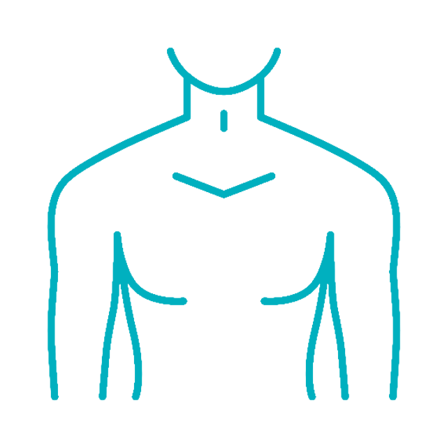
Faster, guideline-compliant and consistent contours for high-precision radiotherapy.
Contour+ is an AI-powered auto-segmentation software that delivers fast and precise automatic delineation of organs-at-risk and lymph node areas. By adhering to industry best practices, Contour+ ensures standardized and consistent contours, resulting in safe and accurate treatment plans delivered with exceptional efficiency.
With Contour+, all users across different clinics begin with the same guideline-based foundation, which facilitates easier inter-clinic collaboration and streamlines research processes.


Contour+ supports 300+ structures for CT and MR, spanning across the entire body, making contouring a matter of just a few minutes. The contours come in pre-defined models that can be further configured to fit within each clinic’s protocol.


Contour+ is GDPR & HIPAA compliant, ensuring that your clinic maintains the highest standards of data protection and patient privacy. All images are pseudo-anonymized and delivered to the MVision AI cloud only to generate the contouring results. Data is uploaded to the cloud through an encrypted connection to be processed by the AI algorithm.
Once the regions of interest (ROIs) are auto-segmented, they are sent back to the clinic via the same encryption/decryption process. Received data is deleted from the cloud server within 24 hours. The entire process, including uploading, processing, and receiving results, takes only a few minutes and happens automatically in the background. This efficiency enables clinicians to make timely, informed decisions, enhancing the overall treatment workflow.
Data Controller:
The clinic is designated as the data controller, retaining full control over patient data while ensuring compliance with privacy regulations.
Data Processor:
MVision AI acts as the data processor, adhering to a comprehensive data processing agreement that guarantees all data handling is secure and compliant with industry standards.


Contour+ is delivered as part of the Workspace+ platform, operating alongside Dose+, Image+, and Adapt+ in one secure, modular environment. Together, these modules support streamlined and standardised radiotherapy preparation workflows.
Contour+ is also delivered as part of GBS™, the guideline-based segmentation solution. Within GBS™, it is combined with Guide and Verify in one comprehensive solution. Together, they support high-quality, guideline-compliant contours and contour assessment.


